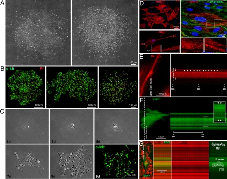
Fig. 3.
In vitro properties of hCSCs. (A–C) Clones formed by hCSCs (c-kit, green) isolated by enzymatic digestion (A and C) or primary explant (B). The number of cells increased with time (C). (D) hCSCs generate myocytes positive for cardiac myosin heavy-chain (MHC), α-SA, and α-cardiac-actinin (α-actinin). Sarcomeres are apparent (Insets); phalloidin, green. (E) Myocyte shortening in cells derived from clonogenic hCSCs was recorded by two-photon microscopy and laser line-scan imaging (Left). The line scan is shown (Right), and arrowheads point to individual contractions. (F) Myocytes derived from EGFP-positive hCSCs, cocultured with neonatal myocytes. EGFP-positive human myocytes shorten (arrowheads) with electrical stimulation. (G) Calcium transients in EGFP-positive human myocytes and EGFP-negative rat myocytes (calcium indicator Rhod-2, red).

