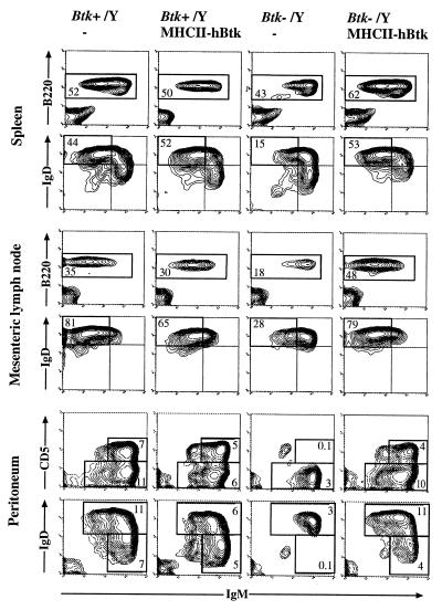
Figure 2.
The effect of the MHCII–hBtk transgene on peripheral B lymphocytes. Three-color flow cytometric analysis of spleen, MLN, and peritoneum from 4-month-old normal and transgenic Btk+/Y males, as well as nontransgenic and transgenic Btk−/Y males. Spleen and MLN cell suspensions were stained with biotinylated anti-IgM and streptavidin-TriColor, anti-IgD-PE, and anti-B220-fluorescein isothiocyanate. (Top) Percentages of cells displayed that are B220+/IgM+ are indicated. (Bottom) Percentages of gated B220+ cells displayed that are IgMlowIgDhigh (fraction I; refs. 14 and 15) are indicated. Peritoneal cells were stained with anti-B220, anti-IgM, and either anti-CD5-PE or anti-IgD-PE; B220+ cells are displayed. The percentages of total cells (including peritoneal macrophages) that are CD5+ (and IgDlow) B cells or conventional (CD5− and IgDhigh) B cells are given. Data are shown as 5% probability contour plots representative of the mice examined; dead cells were gated out, based on forward and side scatter characteristics.

