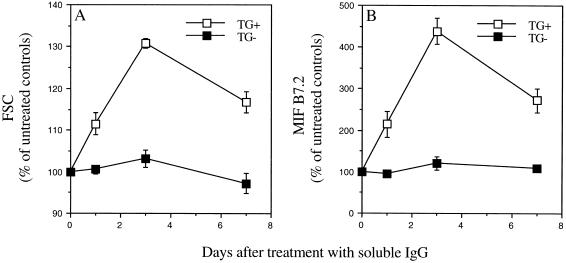
Figure 1.
Loss of hIgM RF-B cells is preceded by activation. AB29 mice were injected i.p. with 2 mg DHGG. Spleen cells were removed 1, 3, or 7 days after treatment, stained for surface expression of B220 (total B cells), hIgM, and the activation antigen B7.2 and then analyzed on the cytofluorograph. (A) Forward scatter (FSC) values of hIgM+/B220+ cells (TG+, □) and hIgM−/B220+ cells (TG−, ▪) from the same mice, as a measure of size. (B) Mean intensity of fluorescence (MIF) of B7.2 staining for hIgM+/B220+ cells (TG+, □) and hIgM−/B220+ cells (TG−, ▪) from the same mice, as a measure of B cell activation. Data shown are means ± SE for groups of three or four mice and represent a percentage of control levels seen on untreated AB29 mice. All time points were significantly different from controls for TG+ cells, no time points were significantly different from controls for TG− cells; P < 0.05.

