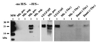
Figure 6.
Western blotting of conditioned medium from megakaryocytic cells. Without heparin-Sepharose enrichment (no H/S), no VEGF could be detected in the conditioned medium (72-h incubation) of CD41a+ (Mk. pos.) and CD41a− (Mk. neg.) cells. Two bands at 21–22 and 17–18 kDa were visible after enrichment of supernatant from CD41a+ cells with heparin-Sepharose (H/S), corresponding to glycolysated and nonglycolysated VEGF165, while no detectable protein was found in the conditioned medium from CD41a− cells. The identity of the bands was confirmed by the positive control (recombinant VEGF165). Thirty minutes after stimulation with thrombin, VEGF165 was also detectable in the supernatant from ex vivo-generated megakaryocytes (Mk. +Thr.) and Dami-cells (Dami +Thr.) after enrichment with H/S, in contrast to unstimulated cells (−Thr.).

