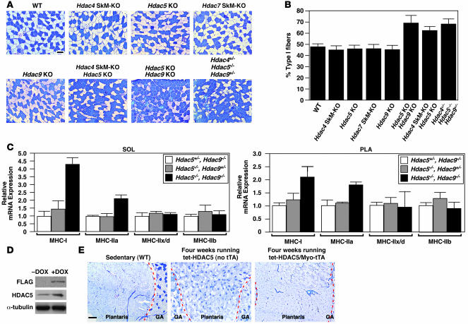
Figure 2. Class II HDACs redundantly regulate slow, oxidative fiber expression.
(A) Soleus muscles from WT, Hdac5–/– (Hdac5 KO), Hdac9–/– (Hdac9 KO), Hdac4fl/–;Myo-Cre (Hdac4 SkM-KO), Hdac7fl/–;Myo-Cre(Hdac7 SkM-KO), and class II HDAC compound mutant mice were analyzed by metachromatic ATPase staining. Type I fibers stain dark blue. Type II fibers stain light blue. Original magnification, ×10. Scale bar: 100 μm. (B) Quantification of fiber-type distribution based on fiber-type analysis in A. (C) Transcripts of MHC isoforms were determined in soleus and PLA muscles from mice of the indicated genotypes by quantitative real-time PCR. (D) Anti-FLAG M2 antibody on a Western blot analysis of proteins isolated from GP muscles of 4-week-old Myo-tTA/tet-HDAC5 mice treated with DOX or 10 days after removal of DOX. Tubulin served as a loading control. (E) Metachromatic ATPase staining of GP muscles harvested from sedentary WT, 4-week-exercised control (tet-HDAC5 [no tTA]), and 4-week-exercised HDAC5 transgenic (Myo-tTA/tet-HDAC5) mice. Original magnification, ×4. Scale bar: 300 μm. Dashed red lines delineate gastrocnemius (GA) muscle from PLA.

