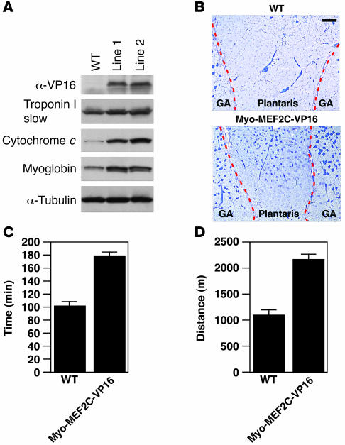
Figure 4. Activated MEF2 is sufficient to increase slow-fiber expression.
(A) Western blot analysis of Myo-MEF2C-VP16 transgene expression using an anti-VP16 antibody. Expression of the slow-fiber–specific troponin I and oxidative markers myoglobin and cytochrome c in protein extract of GP muscles of Myo-MEF2C-VP16 transgenic mice. (B) Metachromatic ATPase staining of gastrocnemius and PLA muscles of WT and Myo-MEF2C-VP16 transgenic mice. Original magnification, ×4. Scale bar: 300 μm. Dashed red lines delineate gastrocnemius muscle from PLA. (C and D) Exercise endurance and muscle performance showing total time running (min; C) and total distance run (m; D) of Myo-MEF2-VP16 transgenic muscles were analyzed by forced treadmill exercise. Eight-week-old Myo-MEF2C-VP16 transgenic and WT male mice with similar body weights were subjected to forced treadmill exercise (n = 5 for each group) on a 10% incline.

