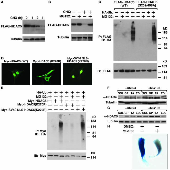
Figure 5. Ubiquitination and degradation of class II HDACs in vitro and in vivo.
(A) C2C12 cells stably expressing FLAG-tagged HDAC5 (C2C12-HDAC5) were treated with cycloheximide (CHX, 25 μM) for 0, 1, 2, or 4 hours before cells were lysed and FLAG-HDAC5 expression was measured by Western blot analysis using anti-FLAG M2 antibody. Tubulin immunoblot showed equivalent loading of each lane. (B) C2C12-HDAC5 cells were treated with cycloheximide and MG132, singly or in combination for 4 hours, and FLAG-HDAC5 expression was analyzed. (C) C2C12-HDAC5 and C2C12 cells stably expressing CaMK-resistant HDAC5 (S259/498A) were transfected with or without HA-tagged ubiquitin (HA-Ub) and treated with or without MG-132 (25 μM) for 4 hours; the ubiquitination status of WT and mutant HDAC5 was analyzed. FLAG expression in inputs shows equal loading. (D) Subcellular localization of Myc-HDAC5 (WT), Myc-HDAC5(K270R), or Myc-SV40 NLS-HDAC5(K270R) in C2C12 cells. (E) The ubiquitination status of cytoplasmic HDAC5 [Myc-HDAC5(K270R)] or nuclear HDAC5 [Myc-SV40 NLS-HDAC5(K270R)] was analyzed. (F and G) WT C57BL/6 males (8 weeks old) were IP injected with DMSO or MG132 for 6 hours. Protein was isolated from SOL, GP, tibialis anterior (TA), and EDL muscles and analyzed for expression of (F) HDAC4 or (G) HDAC5. Tubulin shows equal loading. (H) Treatment with MG132 decreases MEF2 activation. Six hours after DesMEF-lacZ mice were injected with DMSO or MG132, mice were run for approximately 3 hours using forced treadmill exercise. Skeletal muscles were then isolated from DMSO- and MG132-treated DesMEF mice and analyzed for lacZ expression. LacZ expression was reduced in MG132-treated muscles (where class II HDAC expression was increased).

