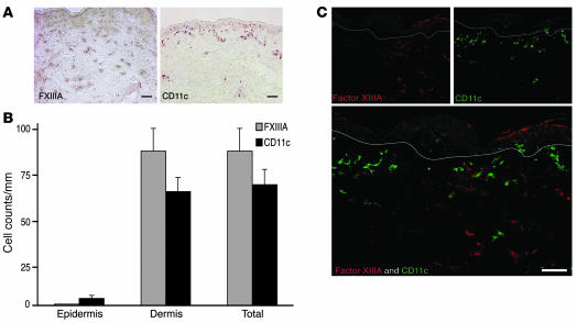
Figure 1. FXIIIA+ and CD11c+ cells are unique dermal populations.
(A) Immunohistochemistry on normal human skin using FXIIIA (left panel) and CD11c (right panel) antibodies (n = 15). FXIIIA+ cells were spread throughout the dermis, while CD11c+ cells were mainly localized to the superficial dermis. (B) There were similar numbers of CD11c+ and FXIIIA+ cells per mm in normal dermis. Error bars indicate SEM. (C) FXIIIA and CD11c identified 2 discrete populations. White lines denote dermo-epidermal junction. Scale bars: 100 μm.

