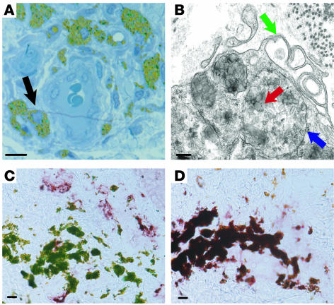
Figure 9. CD163+ cells phagocytose large particles and have the structural features of macrophages.
(A) Tattoo skin section (0.5 μm) stained with toludine blue. Cells containing green tattoo dye in their cytoplasm (black arrow) surrounded a blood vessel. (B) Electron microscopy of a tattoo showed that dye particles (red arrow) were membrane bound (blue arrow) within the cytoplasm of a cell with multiple microvillus protrusions (green arrow). (C and D) Cells containing green tattoo dye particles stained for CD163 (D) but not BDCA-1 (C). Scale bar: 10 μm (A, C, and D); 200 nm (B).

