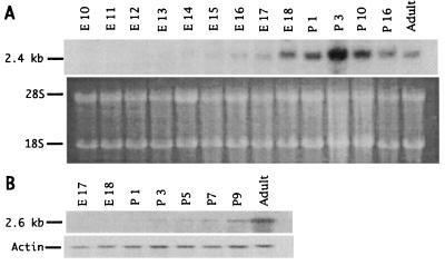
Figure 4.
Temporal patterns of appearance of NPAS1 (A) and NPAS2 (B) mRNA in developing mice. (A Top) Northern blot image developed using a hybridization probe specific to NPAS1 mRNA. (A Bottom) Ethidium bromide staining pattern of RNA samples used for Northern blotting. (B Top) Northern blot image developed using a hybridization probe specific to NPAS2 mRNA. (B Bottom) Image developed using a probe specific for a β-actin mRNA.

