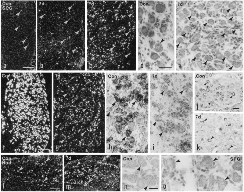
Figure 3.
Dark- (a–c, f, g, l, and m) and bright- (d, e, h–k, n, and o) field micrographs of control (a, d, f, h, j, l, n, and o) and ipsilateral (b, c, e, g, i, k, and m) SCGs (a–k), NGs (l–n), and SPGs (o) of rats, hybridized with probes for Y2R (a–e and l–n), Y1R (o), and NPY (f–i) mRNAs, or immunolabeled for NPY (j and k) 2 (b) or 7 (c, e, g, i, k, and m) days after unilateral transection of both carotid nerves. Few neurons contain Y2R mRNA in normal SCG (arrowheads in a and d) with a small increase 2 days (arrowheads in b), and a marked increase 7 days after Axo (arrowheads in c and e). Most neurons in normal SCGs contain NPY mRNA (arrowheads in f and h) and NPY-like immunoreactivity (arrowheads in j). The intensity of labeling of NPY mRNA is decreased in the neurons (arrowheads in g and i) in ipsilateral SCG 7 days after Axo. The number of NPY+ neurons in ipsilateral SCG is markedly reduced (arrowheads in k). Many neurons in a normal NG express Y2R mRNA (arrowheads in l and n). More NG neurons are Y2R mRNA+ 7 days after Axo (arrowheads in m). Y1R mRNA is expressed in SPG neurons (arrowheads in o). (Bars = 200 μm in a–c, f, g, l, and m; 50 μm in d, e, and h–l; and 20 μm in n and o.)

