Figure 3.
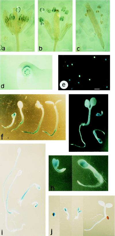
Histochemical localization of GUS activity in plants transgenic for the At-RAB2–GUS fusion. (a) Young flower of line 47–9; 2.1 showing staining of the developing pollen grains in the four most developmentally advanced anthers. The two less advanced anthers lying beneath these four show no staining. (b) Older flower, post anthesis, in which pollen grains in all six anthers are stained. Pollen can be seen on the stigma. (c) Flower from an untransformed plant stained in parallel to those shown in a and b. (a–c, bar = 0.6 mm.) (d) Single flower cut transversely and photographed from above the cut surface, showing that staining is restricted to the anthers even when the other organs are cut to allow better substrate access. (e) Pollen washed out of the anthers of a transgenic plant before staining, and apparently segregating for GUS activity. (Bar = 100 μm.) (f and g) Seedlings of lines 47–9; 2.1 and 47–9; 2.3, respectively, at various stages of development. (h) Enlargments of the two youngest seedlings shown in g. (i and j) Dark-grown and light-grown seedlings of line 47–9; 2.3, respectively, photographed at the same magnification to allow direct comparison of root, hypocotyl, and cotyledon growth and staining.

