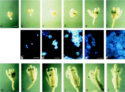
Figure 5.
An inflorescence of a plant from line 47–9; 2.3 was stained for GUS activity for a prolonged period (20 hr) to enable the earliest signs of activity to be visualized. The younger buds were artificially opened to expose the interior, and each bud was photographed. (a–i; bars = 0.5 mm.) Buds were then incubated with the fluorescent DNA stain DAPI, a single anther was excised, and squash preparations from buds a–f were photographed under UV illumination, which revealed the gametophytic nuclei as bright blue fluorescent spots (b′–f′ below the corresponding bud; bars = 30 μm). (a) Apical buds left on the inflorescence axis and not examined individually. (b) Early mononucleate microspores; unstained. (c) Late mononucleate microspores; unstained. (d) Late mononucleate microspores; first signs of GUS activity. (e) Microspore mitosis has occurred (pollen grains show a condensed, brightly fluorescent generative nucleus as well as a larger, more diffuse vegetative nucleus); staining more pronounced. (f) Pollen mitosis has occurred (trinucleate pollen grains with two condensed generative nuclei that are readily visible, and a diffuse vegetative nucleus that is often hard to discern). (g–l) Further development of the flower. Staining reaches its maximum intensity in bud i. (j–l) Staining of the anther appears to wane as they shed pollen; stained pollen appears on the otherwise unstained stigma, and the vascular strand of the filament becomes stained.

