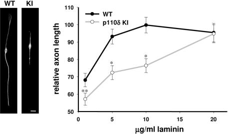
Figure 3. Reduced outgrowth of p110δ KI DRG neuron under limiting substrate conditions.
Left panel, example of E13.5 DRG neurons derived from WT and p110δ KI mice, cultured on 10 µg/ml laminin for 24 h. Right panel, relative axonal length in p110δ KI and WT DRG neurons at 1, 5, 10 and 20 µg/ml laminin (right). Length was expressed relative to that of WT DRG neurons cultured in the presence of 10 µg/ml laminin. Each point represents the mean of at least 3 experiments±SEM, each experiment was carried out in duplicate. n = 60 neurons in each treatment. *p<0.01.

