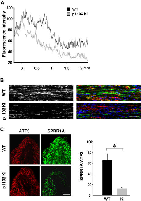
Figure 4. Reduced axonal regeneration in the injured sciatic nerve of p110δ KI mice.
3 days post-injury, the sciatic nerves of WT and p110δ KI mice were fixed and cryo-sectioned. (A) Average relative fluorescence intensity profile of anti-βIII-tubulin labeling across a one-pixel line along the entire nerve segment, following cropping of the micrographs to a fixed pixel segment. (B) High-power micrographs of the sciatic nerve segments 2 mm distal to the injury, labeled with anti-βIII-tubulin (left panels and green in right panels), anti-F4/80 (red) and Hoechst (blue). Scale bar, 50 µm. (C) At 7 days post injury, L4 DRGs of WT and p110δ KI mice were fixed, vibratome-sectioned, and co-labeled with ATF3 (red) and SPRR1A (green). Percentage of SPRR1A and ATF3 co-labeling over ATF3-only positive DRG neurons in WT and p110δ KI mice. Data are from 3 WT and 4 p110δ KI mice, and are presented as mean±SEM. *p<0.05. Scale bar, 50 µm.

