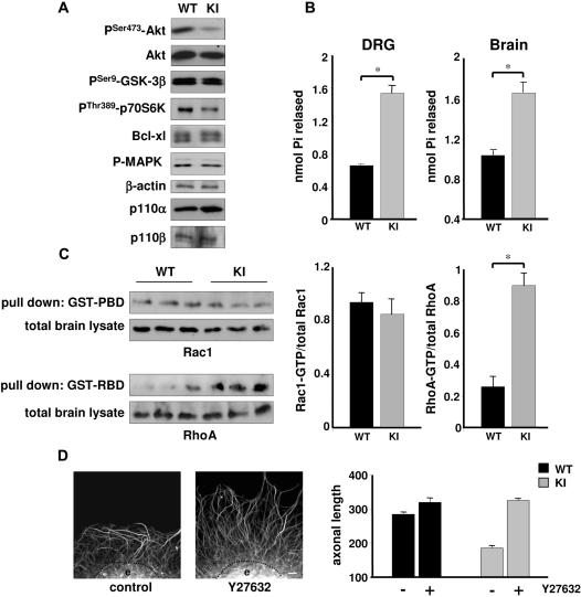
Figure 6. Reduced Akt and increased RhoA/PTEN signaling in DRG neurons with inactive p110δ.
(Α) E13.5 DRG neurons of WT and p110δ KI mice were cultured for 24 h before processing for Western blot analysis using the indicated antibodies. (B) Inactivation of p110δ in p110δ KI mice leads to an increase in lipid phosphatase activity in PTEN immunoprecipitates from homogenates of brain (left panel) or DRGs (right panel) from WT and p110δ KI mice. Graphs show the lipid phosphatase activity in 3 independent experiments, measured in triplicates. p<0.001. (C) Inactivation of p110δ does not affect Rac, but increases RhoA activity. Left panel, brain lysate from WT and p110δ KI mice was subjected to pull-down with GST-PBD or GST-RBD, followed by SDS-PAGE and immunoblotting using antibodies to Rac1 or RhoA. Right panel, graphs represent the mean±SEM of Rac1-GTP (left) or RhoA-GTP loading of 2 experiments, each performed in triplicate. (D) Inhibition of ROCK rescues the effects of p110δ inactivation on axonal elongation at 10 µg/ml laminin. Left panels, example of an E13.5 DRG explant (e) of p110δ KI mice cultured for 24 h in the absence (control) or presence of Y27632 (10 µM). Scale bar, 20 µm. Right panels, length of axons extending from WT and p110δ KI DRG explants, in the absence (−) or presence (+) of Y27632 (10 µM). Each data point represents the mean±SEM of 3 independent experiments.

