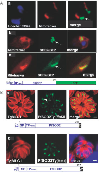
Figure 5. PfSOD2 Is Targeted to the Apicoplast in the Intra-Erythrocytic Stages of P. falciparum .
(A) IFA on live erytrocytes infected with transgenic P. falciparum parasites expressing PfSOD2GFP. The construct starts either at the second methionine ( MNLKIL…) and is controlled by the PfCRT promoter (panel a), or at the first methionine (MLYKLY…) and is controlled by the PfSOD2 promoter (panel b) (see Figure 1A). The mitochondrion is labeled in red with MitoTracker and the localization PfSOD2-GFP to the apicoplast is detected in green. The same construct was stably expressed in the strain 3D7 and the same results were observed. Moreover, PfSOD2 was also shown to localize to the apicoplast only in the gametocytes (panel c). The arrows indicate the apicoplast.
(B) IFA on stable transgenic T. gondii parasites expressing PfSOD2 with a Ty tag at the C-terminus. The construct starts either at the second methionine (panel a) or at the first methionine (panel b). The periphery of T. gondii parasites is labeled in red with anti-MLC1 antibodies, while the localization of the fusion protein PfSOD2-Ty to the apicoplast is shown in green.

