
Fig. 6.
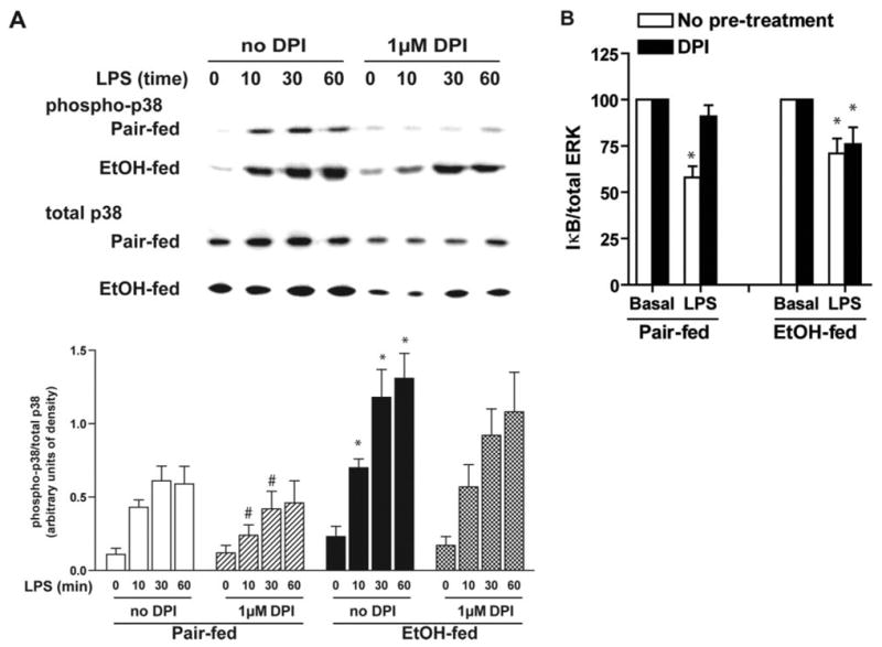
DPI chloride does not suppress LPS-stimulated p38 phosphorylation (A) or IκBα degradation (B) in Kupffer cells from ethanol-fed rats. Kupffer cells from pair- and ethanol-fed rats were cultured 16–18 h. Cells were then preincubated with or without 1 μM DPI for 2 h. (A) Phosphorylation of p38 was measured in response to treatment with 100 ng/ml LPS for 0–60 min. (B) Quantity of IκB-α was assessed by Western blot after 30 min stimulation with or without 100 ng/ml LPS. Values represent means ± sem; n = 7 for p38, and n = 6 for IκB-α. (A) *, P < 0.05, compared with pair-fed; #, P < 0.05, compared with cells not treated with DPI (within a diet group). (B) *, P < 0.05, compared with basal values within each diet group.