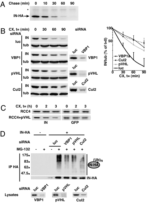
Fig. 4.
Prefoldin and Cul2/VHL complexes are involved in the ubiquitin–proteasome-dependent degradation of IN. (A) IN-HA cells were pulse-labeled with [35S]methionine/cysteine and subsequently chased for indicated time periods prior to immunoprecipitation by using anti-HA antibody. Immunoprecipitated proteins were analyzed by SDS/PAGE and fluorography. IN-HA and a stable contaminating protein (*) reflecting loading are visualized. Quantifications of two independent experiments resulted in an estimated IN half-life of 11 min. (B) IN-HA cells were transfected with siRNA specifically directed against VBP1 (VBP1a), pVHL, or Cul2 or with control luciferase (luc)-directed siRNA. Cells were subsequently treated with the protein synthesis inhibitor cycloheximide (100 μg/ml) for the indicated periods of time prior to lysis, and analyzed by Western blotting with anti-HA antibody and anti-α-tubulin antibody as an internal control. The effect of siRNAs on protein expression was monitored with specific antibodies. Chemiluminescence of the blots was acquired with a Fuji CCD camera (Kanagawa, Japan). For each condition, the IN-HA chemiluminescence signal was quantified by using Image Gauge software and normalized to the α-tubulin signal. Results from five to seven independent experiments are represented on the right. (C) pVHL-negative RCC4 cells stably transfected with pVHL (RCC4+pVHL) or not (RCC4) were transiently cotransfected with IN-HA and GFP expression plasmids. Cells were subsequently treated with cycloheximide (100 μg/ml) for the indicated times prior to lysis. Equal amounts of total protein lysates were then analyzed by Western blotting with anti-HA and anti-GFP antibodies. Chemiluminescence of the blots was quantified as in B, and the IN-HA signal was normalized to the GFP transfection control signal. (D) IN-HA or HeLa cells were transfected indicated siRNAs and subsequently treated with the proteasome inhibitor MG-132 (20 μM) or DMSO prior to lysis. Equal amounts of total cellular proteins were immunoprecipitated by using anti-HA antibody. Immunoprecipitated proteins were analyzed by Western blotting with anti-HA or anti-ubiquitin antibody (IP HA). VBP1 expression in cell lysates was monitored by anti-VBP1 immunoblotting (lysates).

