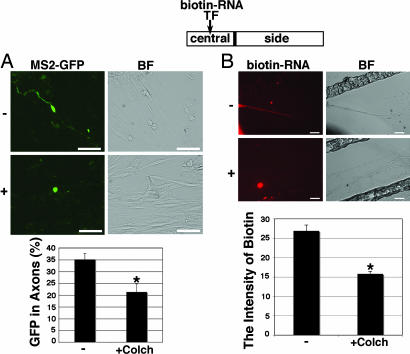
Fig. 4.
kor mRNA transport is microtubule dependent. (A) Micrographs show the mobilization of MS2–GFP-tagged kor mRNA (Left) and the negative control (Right) in DRG neurons with (Lower) or without (Upper) the microtubule disruption drug colchicines. (B) Micrographs show mobilization of biotin-labeled kor mRNA (Left) from the central to the side chambers of the Campenot cultures with (Lower) or without (Upper) colchicines after Copb1 transfection for 24 h to stimulate the transport. Only the side chambers are shown. GFP signals are in green, and biotin-RNA signals are in red. (Scale bars, 25 μm.) Statistical analyses is shown in the graphs (for both, n = 3; *, P < 0.025 for comparing with the control). BF, bright field.

