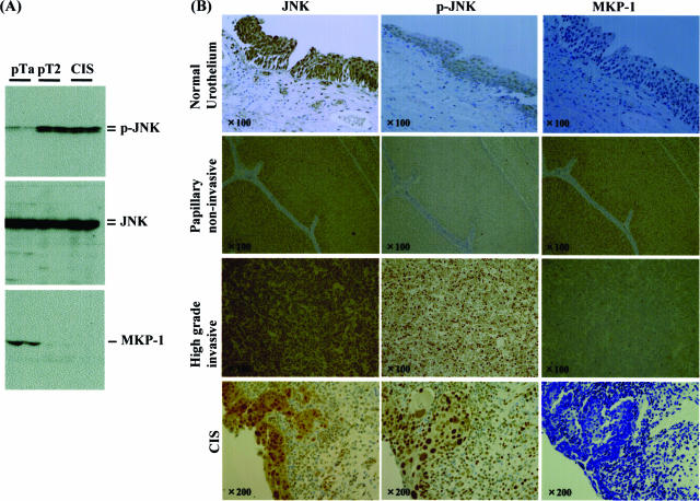
Figure 5.
Immunohistochemical findings for JNK, phosphorylated JNK, and MKP-1 in human urothelial carcinomas of the urinary bladder. A: Tissue samples of urothelial carcinoma obtained by transurethral resection or radical cystectomy were divided into three groups, low-grade (G1, 2)/noninvasive (pTa, pT1) phenotype, high-grade (G3)/invasive (more than pT2) phenotype, and G3/carcinoma in situ. Two cases for each were collected and lysed and then immunoblotting using anti-JNK, anti-phosphorylated JNK, and anti-MKP-1 antibodies was performed. B: All samples and normal urothelial cells distant from cancer foci were stained with these antibodies.

