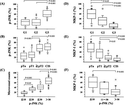
Figure 6.
Box plot figures of immunohistochemical analyses (positive percentages) of human urothelial carcinomas. A, B, D, and E: Relationships between percentages of positive cells for phosphorylated JNK or MKP-1 and grade [G1, 2, and 3)/stage pTis (CIS), pTa, pT1, and ≥pT2]. C: The relationship between microvessel counts and percentages of cells positive for phosphorylated JNK. F: The relationship between percentages of positive cells for MKP-1 and phosphorylated JNK.

