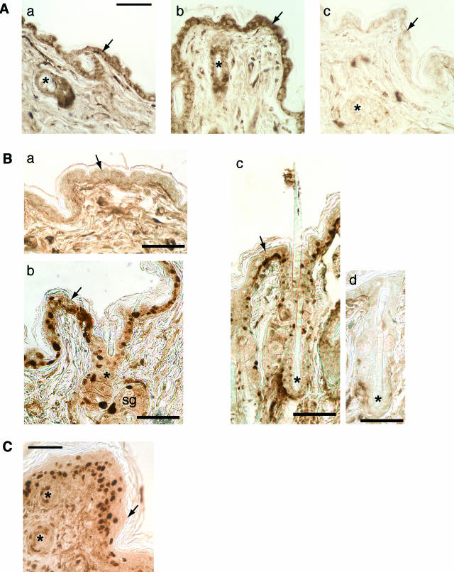
Figure 2.
Immunolocalization of MR in the skin. Paraformaldehyde-fixed sections of the skin of 9-week-old mice (4 weeks after DOX withdrawal) and of E18.5 DT embryos were immunostained with monoclonal anti-MR antibodies. A: Skin from control (a) and DT (b) mice (9 weeks old) immunostained with the 2D6 antibody (endogenous mMR) have comparable epidermal (arrow) and follicular (asterisk) mMR expression over both cytoplasm and nuclei. In c, the 2D6 antibody has been substituted by the unrelated UPC10 mouse antiserum over DT skin (showing low nonspecific signal). B: Immunolocalization of the hMR with the 6G1 antibody (labeling the endogenous MR and the transgenic hMR) in the skin of 9-week-old control (a, without hMR overexpression) and DT (b and c) mice. Cytoplasmic staining was comparable in control (a) and DT (b and c) epidermis. Nuclear signal with 6G1 was found in epidermis (arrow), sebaceous gland (sg), and hair follicle (asterisk) of DT mice (not in control mice). In d, the 6G1 antibody has been substituted by the unrelated UPC10 mouse antiserum over DT skin (showing low nonspecific signal). C: Immunolocalization of the hMR in the skin of E18.5 DT embryos. Nuclear labeling with the 6G1 antibody is apparent over several epidermal layers (arrow) and in hair follicle (asterisk). Bar = 50 μm.

