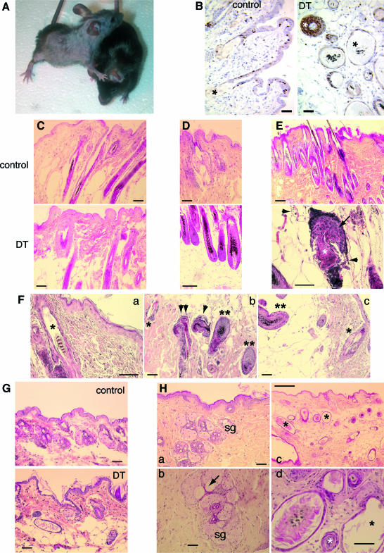
Figure 6.
Cutaneous phenotype of adult control and DT mice. Postnatal expression of hMR results in alopecia and hair follicle cysts. Mice were fed with DOX food from the onset of gestation until the age of 5 weeks, and then DOX was withdrawn to allow MR expression. A: An adult 8-month-old DT mouse (left) with extensive alopecia compared with its control littermate (right). B: Ki 67 staining of the skin of adult (6 months) control and DT mice, showing the increased proliferation in the multilayered hair follicle cysts (white asterisk) in DT mice. Asterisk indicates HF in control and dilated hair follicle cysts in DT mice. Bar = 50 μm. C–H: Consecutive biopsies of murine skin at different time intervals after DOX withdrawal (on week 5 after birth), illustrating the progressive appearance of hair follicle abnormalities and cystic hair follicle degeneration. Lag time after DOX withdrawal: C, 2 weeks; D, 4 weeks; E, 9 weeks; F, 12 weeks; G, 16 weeks; and H, 22 weeks. Bars: 200 μm (in H, c); 50 μm (in all other panels). C: Two weeks after DOX withdrawal, no abnormalities are visible in DT skin. D: Four weeks after DOX withdrawal, control mice show telogen hair follicles, whereas DT skin shows mature anagen hair follicles, as indicated by their characteristic morphology, location (deep in subcutis), and pigmentation (Supplemental Fig. S2 for further evidence of anagen, available at http://ajp.amjpathol.org), suggesting an abnormality in hair follicle cycling. E: Nine weeks after DOX withdrawal, many microscopic fields of DT skin exhibited signs of severe hair follicle dystrophy [as evident from the presence of ectopic melanin granules, melanin incontinence (arrowheads), and malformed inner (white asterisk) and outer (arrow) root sheaths]. This was seen only very rarely and, when present, in a more discrete manner in control skin of this age group. F: Twelve weeks after DOX withdrawal, the skin of DT mice continued to show signs of hair follicle dystrophy, as evidenced by dilated hair canal (asterisk in a) and abnormal hair follicle (HF) architecture and pigmentation (b) as well as of relative asynchrony of hair follicle cycling, as indicated by the simultaneous presence of telogen and anagen hair follicles next to each other (b and c; *, telogen HF; **, anagen HF; arrowhead, dystrophic anagen HF; double arrowhead, dystrophic telogen/catagen HF). G: Sixteen weeks after DOX withdrawal, dermal cysts of HF were apparent in several DT mice, not in controls. H: Histology of the skin 22 weeks after DOX withdrawal in control [a, sebaceous glands (sg)] and DT (b–d) mice. Cysts of sebaceous glands (b, arrow) and of hair follicle (*, c and d) in DT mice are shown. See Table 1 for comments.

