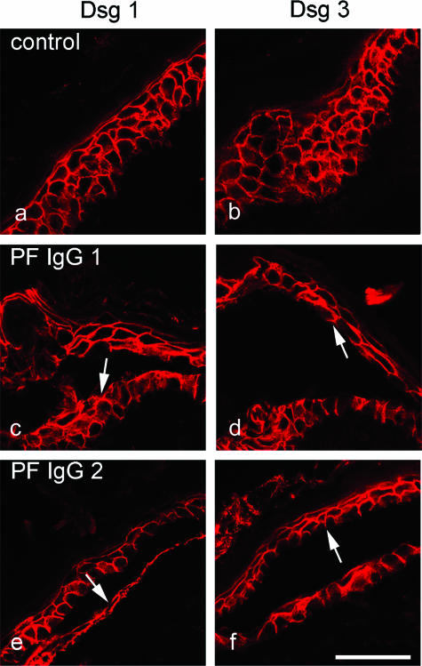
Figure 3.
Similar to PV-IgG, PF-IgG also caused epidermal splitting where both Dsg1 and Dsg3 were present. Skin was immunostained for Dsg 1 (a, c, and e) and Dsg 3 (b, d, and f). Compared with untreated skin (a and b), incubation with PF-IgG containing only Dsg 1-specific antibodies resulted in splitting where expression of Dsg 1 as well as Dsg 3 was found (c–f). Dsg 1 was present underneath the cleavage plane (arrows in c and e), and Dsg 3 immunoreactivity was detected above the split (arrows in d and f). Bar = 50 μm.

