Figure 4.
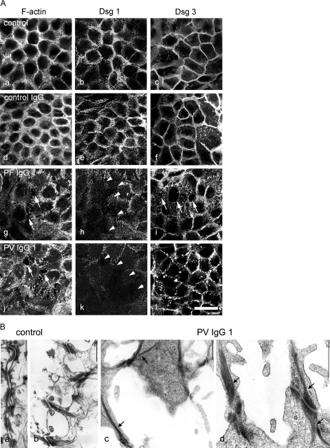
Pemphigus IgG induced cell dissociation in cultured human keratinocytes (HaCaT cells) expressing both Dsg 1 and Dsg 3. A: Cells were stained for F-actin using ALEXA-phalloidin to detect sensitively cell dissociation and to visualize effects on the actin cytoskeleton (a, d, g, and j) and for Dsg 1 (b, e, h, and k) or for Dsg 3 (c, f, i, and l) to label desmosomes. In untreated monolayers (a–c), after incubation with control IgG from a healthy volunteer (d–f), staining of F-actin and Dsg 1 and Dsg 3 was continuous along the cell borders. Incubation with PF-IgG 1 or PV-IgG 1 resulted in keratinocyte dissociation (arrows in g and j) and profound alteration of the actin cytoskeleton and loss of Dsg 1 immunoreactivity in areas of cell dissociation (arrowheads in h and k). After incubation with PF-IgG 1, staining for Dsg 3 was lost at gap margins (arrows in i), whereas PV-IgG 1 resulted in fragmentation of Dsg 3 immunoreactivity, indicating a general loss of desmosomes. Bar = 20 μm. B: The effect of PV-IgG 1 on desmosomes was characterized by transmission electron microscopy (n = 3). In controls (a and b), keratinocyte cell borders were aligned, and numerous desmosomes were visible. After treatment with PV-IgG 1 (c and d), large gaps were present, and desmosomes were reduced in number and restricted to filopodial processes between neighboring cells. These desmosomes were linked to thick keratin filament bundles. Bar = 600 nm.

