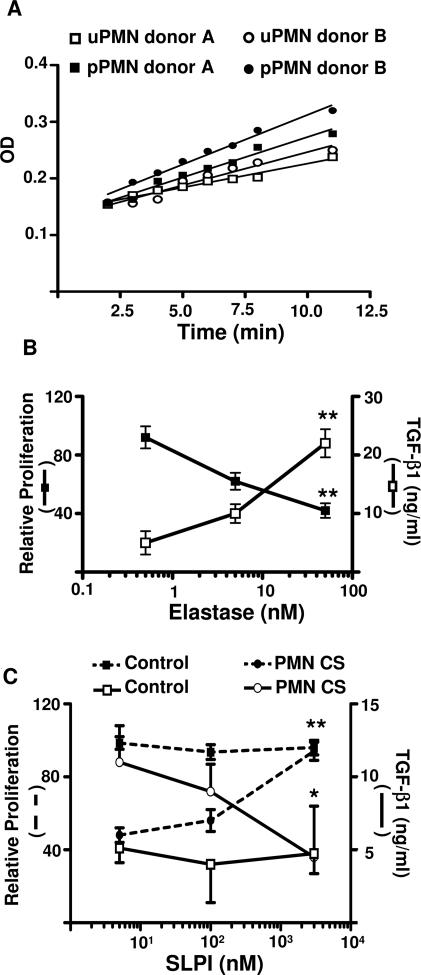
Figure 5.
Effect of elastase on DC lymphocyte allostimulatory ability. A: Elastase activity of PMN CS. PMNs from two healthy donors were primed or not primed with IL-8 for 5 minutes. Afterward, the cells were thoroughly washed and cultured in RPMI-HSA medium for another 3 hours. Then the supernatants were recovered, diluted, and assessed for elastase activity. B: TGF-β production and MLR induced by allogeneic immature DCs previously treated with elastase. Different concentration of elastase was incubated with DCs for 3 hours and then thoroughly washed and cultured in RPMI-HSA medium for 48 hours for assay of TGF-β or incubated with 105 PBMCs for the MLR. Proliferation is depicted relative to the proliferation of MLR cultures induced by untreated DCs (100%). Relative proliferation is depicted as average means of quadruplicates ± SEM from a series of four experiments. *P < 0.05; **P < 0.01 compared with control group, using Dunnett’s multiple comparisons (cpm and TGF-β1 from untreated DCs was 30,739 ± 664 cpm and 3 ± 0.2 ng/ml, respectively). C: To evaluate the effect of SLPI on PMN-CS-treated DC lymphocyte allostimulatory ability and TGF-β1 production, DCs (3 × 10⁴) were incubated for 3 hours with PMN CS and different concentrations of SLPI. Then, cells were thoroughly washed and incubated with 105 PBMCs for the MLR (dotted line) or cultured in RPMI-HSA medium for 48 hours for assay of TGF-β1 production (solid line). Proliferation is depicted relative to the proliferation of MLR cultures induced by untreated DCs (100%). Relative proliferation is depicted as average means of quadruplicates ± SEM from a series of four experiments *P < 0.05 and **P < 0.01 compared with control group, using Dunnett’s multiple comparisons test.

