Figure 2.
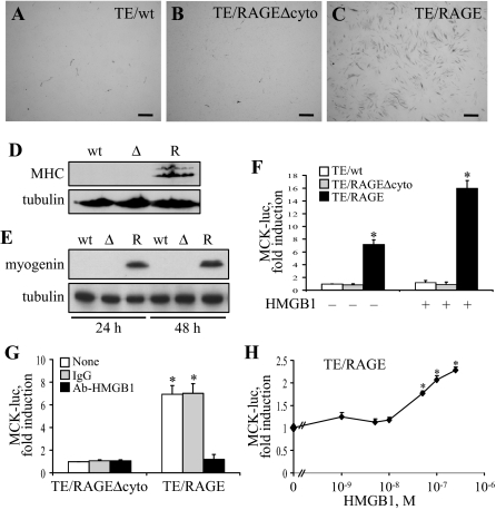
Enforced expression of RAGE in TE671 cells activates the myogenic program on stimulation with HMGB1. A–C: TE671/wt, TE671/RAGEΔcyto, and TE671/RAGE cells were cultivated in DM for 4 days, fixed, and subjected to immunocytochemistry for detection of MHC. D: Same as in A–C except that the cells were solubilized and subjected to Western blotting for detection of MHC. R, wt, and Δ stand for TE671/RAGE, TE671/wt, and TE671/RAGEΔcyto cells, respectively. E: Same as in A–C except that the cells were cultivated in DM for 24 and 48 hours before Western blotting for detection of myogenin. F: TE671/wt, TE671/RAGEΔcyto, and TE671/RAGE cells were transiently transfected with MCK-luc reporter gene, switched to DM, cultivated for 24 hours in the absence or presence of added HMGB1 to 100 nmol/L, and harvested to measure luciferase activity. G: Same as in F except that TE671/RAGEΔcyto and TE671/RAGE cells were cultivated in the presence of either 2.5 μg/ml nonimmune IgG or 2.5 μg/ml anti-HMGB1 antibody for 48 hours. H: Same as in F except that TE671/RAGE cells were cultivated in the presence of added HMGB1 to the concentrations indicated. One representative experiment of three is shown (A–E). Averages of three independent experiments ± SD (F–H). *Significantly different from control (first column from left in F and G; TE671/RAGE cells in the absence of additions in H) (P < 0.01). Bars = 200 μm (A–C).

