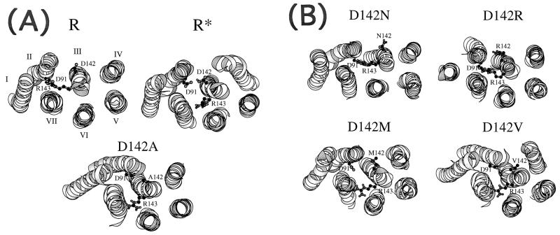
Figure 3.
(A) View of the minimized average structures of wild-type α1B-AR (R and R*) and of the D142A mutant in a direction parallel to the helix main axes from the intracellular side. The R structure carries D142 in the deprotonated (anionic) form, whereas the R* structure carries D142 in the protonated (neutral) form. For clarity, only the α-helical domains have been represented, whereas the loops have been excluded from the picture. (B) View of the minimized average structures of the D142N, D142R, D142M, and D142V mutants in a direction parallel to the helix main axes from the intracellular side.

