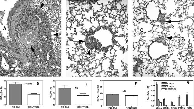
Figure 1.
PH and perivascular inflammation is transient in immunocompetent mice. A: Pulmonary tissue in an immunocompetent mouse 14 days after inoculation with Pneumocystis showing marked perivascular inflammation (arrowhead) and enlarged medial layers of small pulmonary arteries (arrow). B: Similar view at 30 days after infection with limited residual perivascular inflammation and reduced medial hypertrophy in small arteries. C: Comparable view in an uninfected control. D and E: At 14 days after inoculation with Pneumocystis, mean peak right ventricular pressure (RVP) is significantly elevated, whereas the mass of the RV, expressed as the percentage RV mass is of the combined mass of the left ventricle + septum (LV + S), is slightly, but not significantly higher; n = 9 (infected) or 6 (control). F: At 30 days after infection, RVP is no longer significantly elevated [n = 6 (infected) or 7 (control)]. NS, nonsignificant. G: Inflammatory cells in the BALF are greatly reduced at 30 days compared with 14 days (Macs, macrophages; CD4s and CD8s are lymphocytes only; PMNs, neutrophils; EOS, eosinophils). Values are means ± SEM; P is probability that value is statistically equal to control animals. Original magnifications, ×400.

