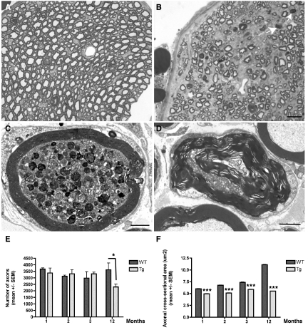
Figure 2.
A and B: Semithin transverse sections of the sciatic nerve of 12-month-old wild-type (A) and Tg30tau (B) mice. There is a loss of myelinated axons in the Tg30tau mouse, and myelin ovoids are observed (arrows in B). C and D: Ultrastructural section of the sciatic nerve of Tg30tau mice. C: An axon shows accumulation of dense and lamellar bodies and degenerating mitochondria in a 3-month-old mouse. D: A degenerating axon is surrounded by a myelin sheath showing Wallerian degeneration in a 12-month-old mouse. E: The mean number of axonal profiles in transversal sections of the sciatic nerve is similar in young adult wild-type and Tg30tau mice but is significantly decreased in aged Tg30tau mice (n = 3 to 5 for wild-type and n = 3 to 6 for Tg30tau mice, at each age) (two-way analysis of variance with Bonferroni post test). F: The mean cross-sectional area of axons in the sciatic nerve is smaller in Tg30tau mice and increases with age in wild-type but not in Tg30tau mice (same animals as in E) (two-way analysis of variance with Bonferroni post test). *P < 0.05, ***P < 0.001. Scale bars: 5 μm (A and B); 1.5 μm (C and D).

