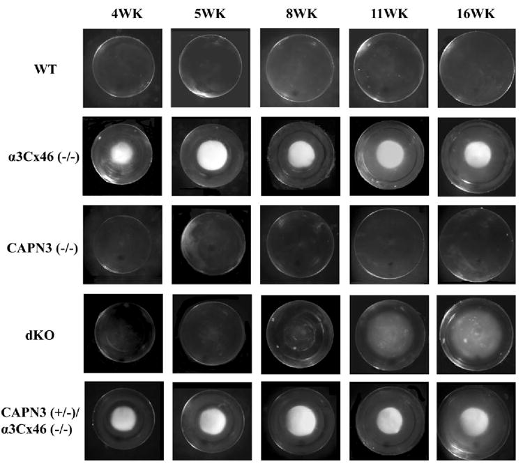
FIGURE 2.

Visual examination of lenses from the different KO mice. Shown are lenses dissected from fresh eyes of WT, α3Cx46−/−, CAPN3−/−, dKO, and CAPN3+/−/α3Cx46−/− mice at the age of 4, 5, 8, 11, and 16 weeks and photographed with a dissection microscope. Note that cataract formation in the dKO mouse lens is delayed and its appearance is changed compared to the α3Cx46−/− mice.
