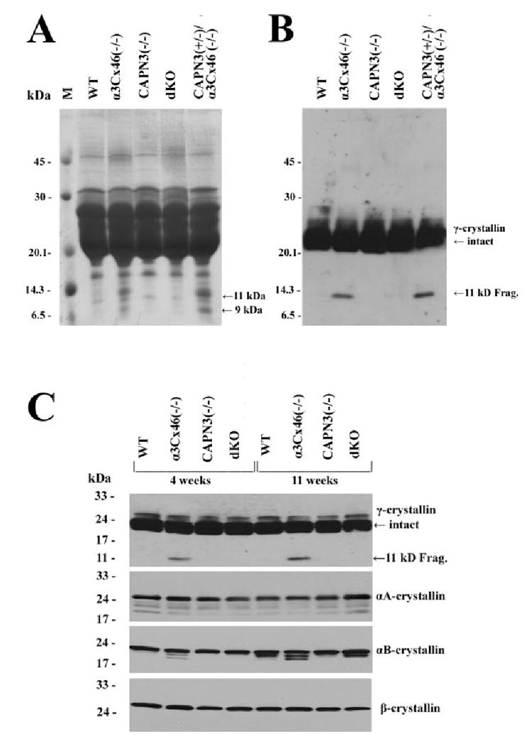
FIGURE 5.

Analysis of crystallins in the lenses from the different mice. (A) A Coomassie blue–stained gel of the total lens homogenate from 11-week-old lenses of WT, α3Cx46−/−, CAPN3−/−, dKO, and CAPN3+/−/α3Cx46−/− mice. Equal amounts of the samples (200 μg) were separated on a 15% SDS-polyacrylamide gel and stained with Coomassie blue. Arrows: the bands (in lanes of α3Cx46−/− or CAPN3+/−/α3Cx46−/− samples) corresponding to the 9- and 11-kDa cleaved forms of γ-crystallins. (B) Western blot analysis of samples used for (A). Equal amounts of the total homogenate samples (5 μg) were subjected to 15% SDS-PAGE and Western blot analysis. An anti-γ-crystallin polyclonal antibody was used. Arrows: position of the intact and cleaved form of γ-crystallin protein. (C) Crystallin profiles in the lenses of the different KO mice. Homogenates from WT, α3Cx46−/−, CAPN3−/−, and dKO lenses were prepared from mice of 4 or 11 weeks of age. Equal amounts of the total homogenate samples (5 μg for γ-crystallins and 10 μg for other crystallins) were subjected to 10% to 20% gradient SDS-PAGE and Western blot analysis. Four different antibodies—anti-γ-, anti-αA-, anti-αB-, and anti-β-crystallins—were used to analyze the samples by Western blot analysis. Arrows: position of the intact and cleaved form of γ-crystallin protein.
