
Figure 6.
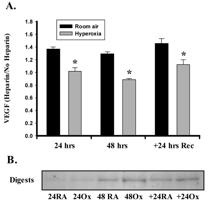
Hyperoxia alters the binding of VEGF to heparin/heparan sulfate moieties. A. A549 cells were exposed to hyperoxia or room air in the presence or absence of 10 μg/ml of sodium heparin. Following the exposure period, medium was removed and assayed for the presence of VEGF by ELISA. Values are expressed as a ratio of medium + heparin/medium alone. Heparin increased soluble VEGF content at each time in room air (RA) but not in hyperoxia (Ox) cells. Columns represent mean (n=3) and bars standard error of the mean. *Denotes p<0.05 of Ox vs. RA at each time point. B. Representative immunoblot showing VEGF protein digested from cells with Heparinase III. Expression was assessed at 24 and 48 hrs of exposure, and following recovery for 24 hrs in room air (+24). Digests were prepared by adjusting Heparinase III concentration to cell number as described in MATERIALS AND METHODS.