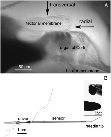
FIGURE 1.
(A) Image of a radial cross section of a gerbil cochlea at a middle-turn location. Shown are the basilar membrane, the organ of Corti, and the tectorial membrane. The osseous spiral lamina can be seen in the lower left side of the image. The arrows indicate the points on the tectorial membrane selected for stiffness measurements and the directions in which the measuring probe was advanced. (B) Image of the sensor system used to measure stiffness. It consists of a solid needle tip (diameter, 25 μm) attached to a piezoelectric “sensor” bimorph, attached in turn to a piezoelectric “driver” bimorph. The insert shows magnified views of the probe tip from the side (inset, lower) and head-on (inset, upper).

