
Figure 2.
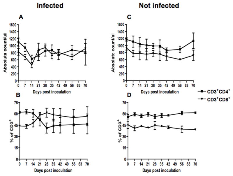
Absolute counts (top) and percentages (bottom) of CD4 and CD8 lymphocytes in peripheral blood of SHIVsf162p3 infected (A & B) (n=10) and uninfected (C & D) (n=3) macaques. Data presented are means ± standard errors of CD4 and CD8 T cells at different time points of infection.