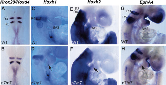
Figure 3.
Hindbrain segmentation and patterning in the Fgfr1n7/n7 mutants. Whole-mount mRNA in situ hybridization analysis of the expression of molecular markers for the hindbrain segmentation in wild-type (A,C,E,G) and Fgfr1n7/n7 mutant (B,D,F,H) embryos. Dorsal views of E8.5 embryos hybridized with the Krox20 and Hoxd4 probes (A,B). Side views of E9.5 embryos hybridized with Hoxb1 (C,D), Hoxb2 (E,F), and EphA4 (G,H) probes. (D,F,H) Arrows indicate affected second branchial arch in mutants. (A–H) Note the similar gene expression patterns in wild-type and Fgfr1n7/n7 embryos. R3–5, rhombomere 3–5; BA2, second branchial arch.

