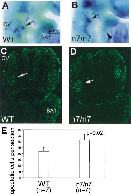
Figure 6.
Analysis of cell death in Fgfr1n7/n7 mutants. Detection of apoptotic cells by Nile blue sulfate (NBS) staining (A,B) and TUNEL staining (C,D) on sections in E9.5 wild-type (A,C) and Fgfr1n7/n7 (B,D) embryos. Cell death, proximally to the second branchial arch (indicated by arrows in A–D), is significantly increased in Fgfr1n7/n7 mutants (B,D). (E) The number of apoptotic mesenchymal cells, counted from sections of the second branchial arch region of wild-type and Fgfr1n7/n7 embryos, is presented graphically. BA1, first branchial arch; BA2, second branchial arch; OV, otic vesicle.

