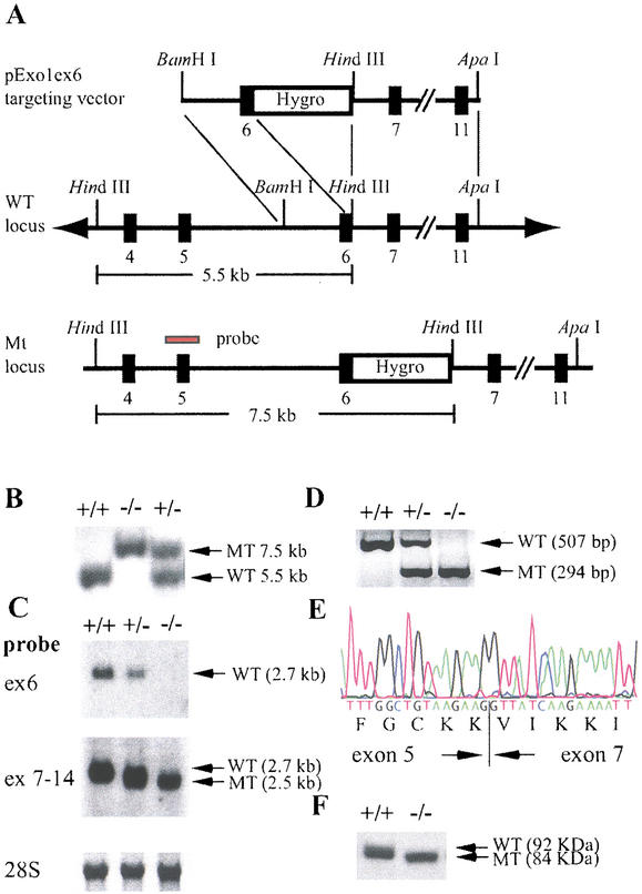
Figure 1.
Disruption of the mouse Exo1 gene by homologous recombination. (A) Map of the targeting vector, the Exo1 wild-type locus, and the modified locus. The probe used for Southern blot analysis is indicated (red bar). (B) Southern blot analysis of HindIII-digested genomic DNA from F2 animals; +/+, wild-type; +/−, heterozygote; −/−, homozygote. (C) Northern blot analysis of poly(A) RNA from Exo1 ES cell lines using exon 6 (ex 6) and exons 7–14 (ex 7–14) probes. (D) RT–PCR analysis of total RNA from Exo1 ES cell lines using primers located in exons 5 and 7. (E) DNA sequence of the mutant RT–PCR product in Exo1−/− mice shown in D. Note that exon 5 is spliced to exon 7 in frame, confirming the deletion of exon 6. (F) Western blot analysis of protein extracts from wild-type and Exo1−/− ES cells using an anti-Exo1 antibody. Note the slightly faster migrating protein species in Exo1−/− cells.

