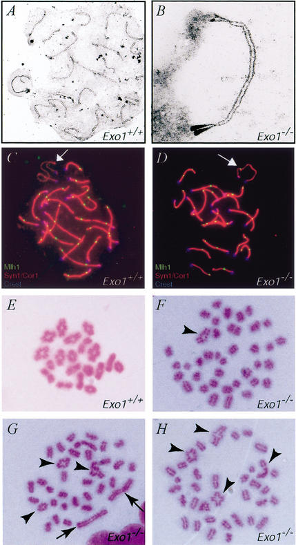
Figure 5.
Chromosome pairing and synapsis during meiosis I. (A,B) electron micrographs of silver-stained chromosome spreads from Exo1+/+ (A) and Exo1−/− (B) spermatocytes, showing normal pairing and synapsis at pachynema. (C,D) Correct spatial and temporal localization of Mlh1 (green, FITC signal) on meiotic chromosomes from Exo1+/+ (C) and Exo1−/− (D) mice stained with a dual antibody against Cor1 and Syn1 and TRITC/red secondary antibody. The centromere is marked with human CREST antisera (blue, Cy5 signal). (E–H) Air-dried chromosome preparations from Exo1+/+ (E) and Exo1−/− (F–H) spermatocytes, showing abnormal metaphase configurations in the absence of Exo1. Although some crossovers remain (arrowheads), the majority of chromosomes are either univalents or appear to be achiasmate bivalents (arrows).

