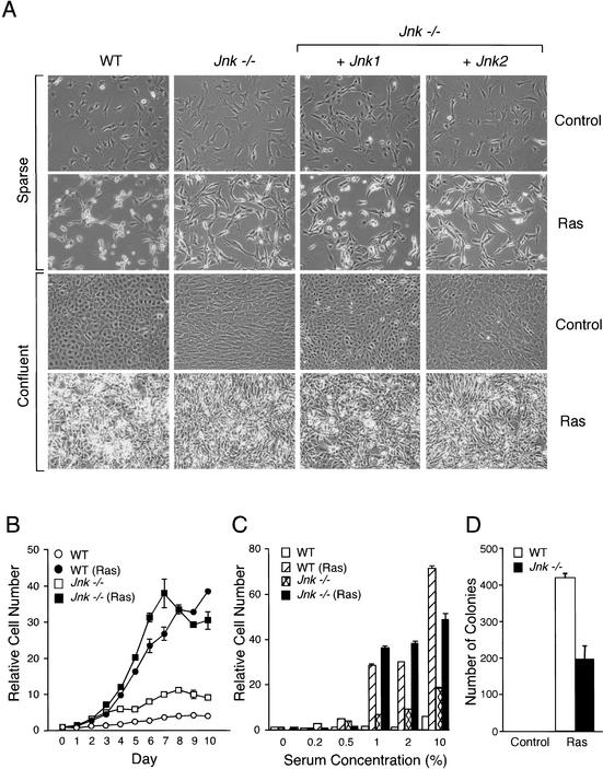
Figure 3.
Analysis of wild-type and Jnk-null fibroblasts transformed with Ras in vitro. (A) Cell morphology in sparse and confluent cultures was examined by phase contrast microscopy. The morphology of wild-type cells, Jnk-null cells, and Jnk-null cells complemented with Jnk1 or Jnk2 is shown. (B) Cell proliferation in medium supplemented with 10% fetal calf serum was examined by crystal violet staining (mean OD at 590 nm ± S.D.; n = 3) following the addition of 1 × 10⁴ cells to 20-mm tissue culture dishes. The data are expressed as relative cell number. (C) The saturation growth density in different concentrations of serum was examined by crystal violet staining (mean OD at 590 nm ± S.D.; n = 3). Relative cell numbers were measured at day 0 (D = 0) and after culture for 10 d (D = 10). (D) Wild-type and Jnk-null fibroblasts were plated in soft agar, then incubated for 14 d, and the number of colonies was measured. The data presented are the mean ± S.D. of triplicate data obtained in three independent experiments.

