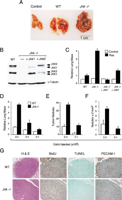
Figure 4.
JNK suppresses tumor development in vivo. (A,B) Wild-type and Jnk-null cells (5 × 10⁵) were injected into the tail vein of 12-week-old male athymic nude mice (Charles River). The mice were injected with 400 μg of BrdU on day 13 and euthanized on day 14. (A) Representative Ras-induced tumor nodules in the lungs are illustrated. (B) Wild-type and Jnk-null cells were examined by immunoblot analysis using antibodies to JNK and α-tubulin. Complementation assays were performed using Jnk-null cells expressing Jnk1 or Jnk2. (C) The lung mass as a percentage of total body mass (mean ± S.D.; n = 5) is presented as relative lung mass. The data presented are representative of three independent experiments. (D–F) Dose-response analysis of tumor formation by Ras-transformed wild-type and Jnk-null cells. The effects of injecting different numbers of transformed cells on the tumor burden (D), the number of tumor nodules (E), and the number of tumor nodules with a surface greater than 2 mm2 (F) are shown. The data presented represent the mean mass ± S.D. (n = 5). (G) The wild-type and Jnk-null tumor nodules were examined by immunocytochemistry. Representative images of sections stained with hematoxylin and eosin (H&E), with an antibody to BrdU to detect proliferating cells, by TUNEL assay for apoptotic cells, and with an antibody to the endothelial cell protein PECAM-1.

