Figure 2.
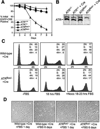
ATRΔ/− cells divide and then exit the cell cycle. (A) Asychronously expanding ATRflox/− MEFs expressing EGFP–Cre are selectively lost from culture. The cells that continue to express EGFP–Cre over the course of 8 d of growth were quantitated as a percentage of their initial representation 1 d after infection with lenti-EGFP–Cre. ▪, wild type; ♦, ATR+/−; ●, ATRflox/+; ▴, ATRflox/−. (B) Western blot quantitation of ATR protein levels in ATRflox/− and ATR+/− MEFs 60 h after NLS-Cre expression (lenti-Cre). (C) Cell cycle analysis of lenti-Cre-infected wild-type and ATRflox/− MEFs. DNA content was determined by propidium iodide staining and flow cytometric analysis. (D) Exit of ATRΔ/− cells from the cell cycle. Cells treated as in C are shown after 1 and 6 d of FBS stimulation. Cells were split 1:4 at day 3.

