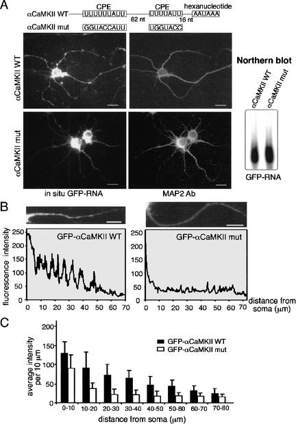
Figure 1.
Facilitation of mRNA transport by the CPE. (A) Hippocampal neurons were infected with Sindbis virus carrying GFP fused to a 170-bp 3′-UTR of wild-type αCaMKII that contains two CPEs, or a mutated αCaMKII 3′-UTR in which the CPEs have been mutated. Seven hours after infection, the neurons were fixed for in situ hybridization with GFP sequences, followed by the MAP2 immunostaining. The Northern blot shows comparable expression of the two constructs in infected neurons. (B) Quantification of dendritic GFP mRNA as assessed by in situ hybridization. The average GFP RNA signal (wild-type or mutant αCaMKII 3′-UTR) in each segment of one major dendrite from each neuron was measured (NIH-Image) and plotted against the distance of the signal from the soma. (C) The mean intensity of each 10-μm dendritic segment was calculated from neurons expressing GFP-αCaMKIIwt (31 dendrites) or GFP-αCaMKIImut (27 dendrites) and presented in histogram form; error bars show S.E.M. The difference in the amount of dendritic GFP hybridization signal between the two RNAs is significant (p < 0.05, Student's t test). Bars, 10 μm.

