Figure 3.
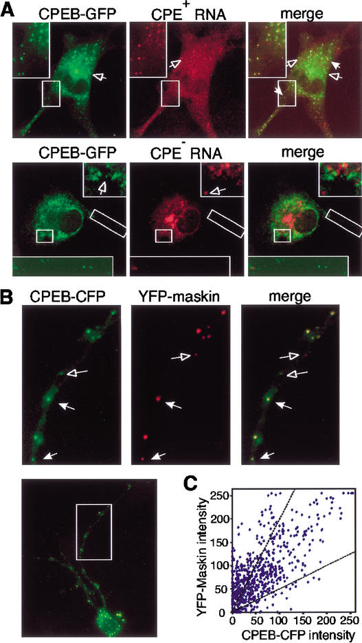
CPEB-GFP forms particles and colocalizes with RNA and maskin. (A) Colocalization of CPEB-GFP and CPE-containing RNA in B104 cells. CPEB-GFP plasmid DNA and Alexa-546-labeled 3CPE-containing or -lacking polylinker RNA were coinjected into B104 cells and imaged by dual-channel confocal microscopy. Particles containing either CPEB-GFP (green) or Alexa-546-labeled RNA (red) are denoted by open arrows; particles containing both CPEB-GFP and RNA (yellow) are denoted by solid arrows. (B) Hippocampal neurons were cotransfected with CPEB-CFP (green) and YFP-maskin (red) and imaged by confocal microscopy. The arrows in the magnified regions denote particles that contain both CPEB-CFP and YFP-maskin proteins. (C) Colocalization of CPEB-CFP and YFP-maskin in 620 RNA particles from dendrites of eight double-transfected cells was quantified by single-particle ratiometric analysis. The two broken lines define the region in which CPEB and maskin in particles are present within 1:2 and 2:1 intensity ratios.

