Figure 7.
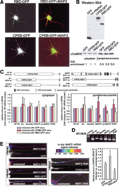
CPEB facilitates RNA transport to dendrites. (A) Infection of hippocampal neurons with Sindbis virus harboring RBD-GFP (RBD refers to the RNA binding region of CPEB) and CPEB-GFP, which were also immunostained with antibody directed against MAP2. Note that whereas CPEB-GFP was detectable in the soma and dendritic arbors, RBD-GFP was confined to the soma. Bars, 10 μm. (B) The extracts from neurons infected with recombinant Sindbis virus were immunoblotted for GFP and CPEB-GFP fusion proteins. The bottom portion of the panel shows the degree of synaptoneurosome enrichment following infection with recombinant virus. Using a densitometric scan of a Western blot probed for αCaMKII, the degree of enrichment was comparable for each preparation. (C) Key features of neurofilament-M (NF-M), Arc, and dendrin mRNAs, which lack obvious CPEs, and MAP2, αCaMKII, and IP3 receptor mRNAs, which contain multiple putative CPEs. Synaptoneurosomes were prepared from GFP-, CPEB-GFP-, and RBD-GFP-containing Sindbis virus-infected cells, and the amount of the RNAs noted above was quantified by real-time PCR. To take into account small variations of the amount of cDNA in each preparation, the amount of RNA in synaptoneurosomes following GFP-expressing virus infection was used as the normalization standard. Error bars indicate the S.E.M., and the asterisks denote significance (p < 0.05, Student's t test). (D) The gel at the bottom shows the final PCR amplification products. (E) In situ hybridization of MAP2 RNA. Hippocampal neurons were infected with virus containing GFP, CPEB-GFP, or RBD-GFP and processed for in situ hybridization for MAP2 RNA. The intensity of the hybridization signal is color-coded. On the same coverslip, the MAP2 RNA hybridization signals were compared between GFP-infected and uninfected neurons, CPEB-GFP-infected and uninfected neurons, and RBD-GFP-infected and uninfected neurons, quantified, and plotted as a ratio of the two (right). The differences between the MAP2 RNA signals in dendrites are significantly different between CPEB-GFP-infected and uninfected neurons, and between RBD-GFP-infected and uninfected neurons (p < 0.05, Student's t test). The GFP fluorescence in dendrites from infected neurons is also shown.

