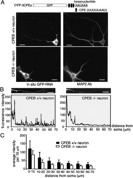
Figure 8.
Deficient transport of CPE-containing RNA in neurons from CPEB knockout mice. (A) Wild-type (CPEB+/+) and knockout (CPEB−/−) hippocampal neurons were infected with Sindbis virus expressing GFP-3CPEs. The neurons were subjected to in situ hybridization for GFP RNA, and also costained with MAP2 antibody. (B) The quantification of the in situ hybridization signal for GFP was performed as in Figure 1B. (C) The mean intensity of each 10-μm dendritic segment was calculated from 20 CPEB+/+ and 20 CPEB−/− neurons expressing GFP-3CPE and presented in histogram form; error bars show the S.E.M. The difference in hybridization signal between the two RNAs is significant (p < 0.05, Student's t test). Bars, 10 μm.

