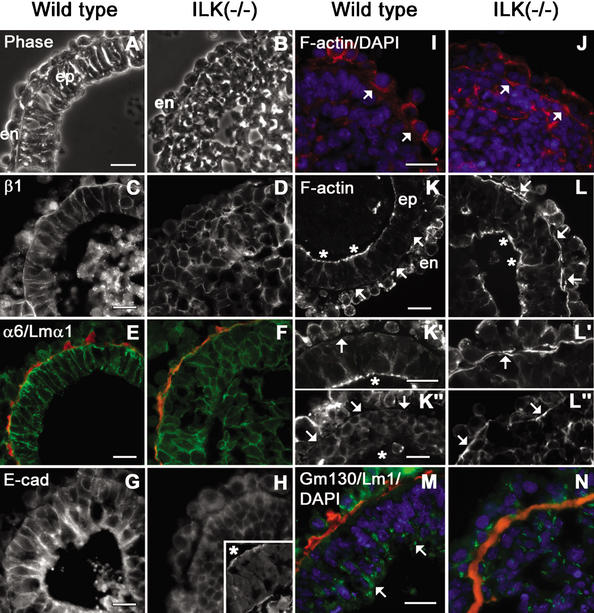
Figure 3.
Localization of laminins, integrins, E-cadherin, β-catenin, and F-actin in EBs. Wild-type and ILK-null EBs were cultured in suspension for 7 d and examined by phase-contrast (A,B) and fluorescence microscopy prepared for immunostaining of β1- and α6-integrin subunits, laminin α1, E-cadherin, F-actin, and GM130 (C–N). (A) Wild-type EBs consisted of an outer layer of endoderm (en), a BM, and pseudostratified columnar epiblasts (ep) facing a sharply demarcated central cavity. (B) The ICM of ILK-null EBs failed to differentiate into epiblasts. (C–F) β1- and α6-integrins were localized at the plasma membrane of epiblast (wild-type) and ICM cells (ILK-null), and to a lesser degree, of the endodermal cells. (E,F) Intense integrins were also seen in the BM zone where laminin 1 (laminin α1 epitope in red) and other BM components (data not shown) were present. (G,H) E-Cadherin was distributed in cell peripheries both in wild-type and ILK-null epiblast cells. (H, inset) The apical distribution of E-cadherin facing the slit-like cavity (indicated by an asterisk) at higher magnification. (I) A weak F-actin signal (F-actin in red; DAPI in blue) was present evenly distributed beneath the plasma membrane of ICM cells adjacent to the BM. The arrow indicates the location of the BM. (J) ICM cells adjacent to the BM in ILK-null EBs show strong F-actin staining prior to polarization. The arrow indicates F-actin beneath the plasma membrane of ICM cells adjacent to the BM. (K) F-Actin was strongly present on an apical belt within epiblast cells that faced the cavities (K,K‘; asterisks) but not in the BM zone (K,K‘,K"; arrows). (L) ILK-null EBs showed strong F-actin staining at the side facing the BM (L,L‘,L"; arrows), although ILK-null EBs with small cavities or slits could gain a columnar shape and F-actin at their apices (L; asterisks). (M,N) GM130 (GM130 epitope in green; laminin 1 epitope in red; DAPI in blue) in wild-type EBs was observed in epiblast cells adjacent to the cavity edge but not ILK-null EBs. α6, α6-integrin subunit; β1, β1-integrin subunit; E-cad, E-cadherin; Lm1, laminin 1; Lmα1, laminin α1. Bars, 20 μm.

