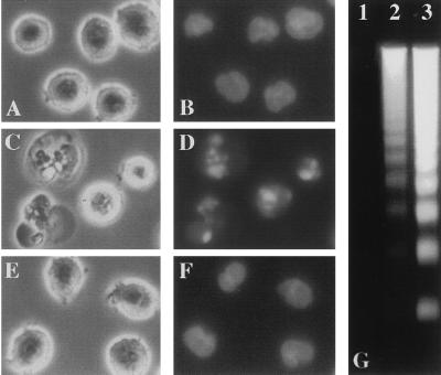
Figure 1.
(A–F) Phase-contrast photomicrographs (A, C, and E) of HL60 cells and photomicrographs of the same cells after nuclear staining with Hoechst 33324 (B, D, and F) viewed 6 h after the following treatments: 0.1% DMSO (A and B), 50 μM PSI (C and D), or 50 μM E64 (E and F). (G) Oligonucleosomal DNA fragmentation from PSI-treated cells: 0.1% DMSO (lane 1), 10 μM PSI (lane 2), or 50 μM PSI (lane 3). Soluble DNA fragments were extracted, separated on a 1.7% agarose gel, and stained with ethidium bromide. The DNA fragmentation patterns were similar in cells treated with TPCK, DCI, LLnL, or LLnV (not shown).

