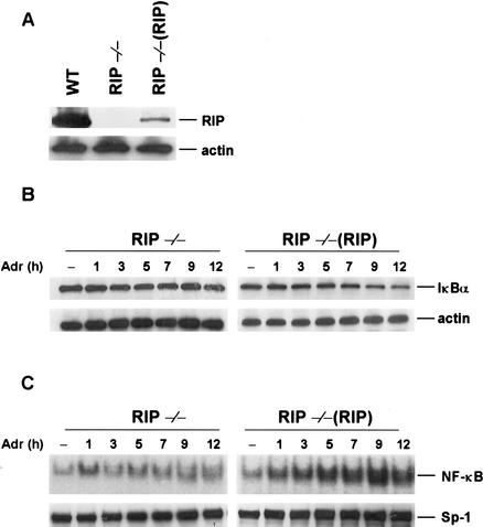
Figure 3.
Expression of RIP reconstituted DNA damage-induced IκBα degradation and NF-κB activity in RIP−/−(RIP) fibroblast cells. (A) The protein expression levels of RIP in wild-type, RIP−/−, and RIP reconstituted stable cell lines of RIP−/− [RIP−/−(RIP)] fibroblasts. The same amount of cell extract from each cell line was applied to SDS-PAGE for Western blotting with anti-RIP and anti-β-actin antibodies. (B) Reconstitution of Adr-induced IκBα degradation in RIP−/−(RIP) cells. Mouse RIP−/− and RIP−/−(RIP) cells were incubated with Adr (10 μg/mL) for various times as indicated. Cell extracts were applied to SDS-PAGE for Western blotting with anti-IκBα and anti-β-actin antibodies. (C) Reconstitution of Adr-induced NF-κB binding activities in RIP−/−(RIP) cells. Mouse RIP−/− and RIP−/−(RIP) cells were incubated with Adr (10 μg/mL) for various times as indicated in the figure. NF-κB activity was measured by EMSA as described in Figure 2.

